Major: Biosciences - Cell Biology and Genetics
Research Advisor: Dr. Kartik Venkatachalam, McGovern Medical School at UTHealth
The common fruit fly, Drosophila, is often a nuisance that we swat away when it nears our food. In the scientific world, however, Drosophila is studied at length. For over a century, Drosophila has been used as a model organism by researchers to study a diverse range of biological processes. One such study done by Abinav Sankaranthi, a senior Biosciences major with a concentration in Cell Biology and Genetics at Rice University, delves into the Drosophila nervous system.
Since the spring semester of his freshman year, Abinav has been working with Dr. Kartik Venkatachalam at McGovern Medical School at UTHealth. Currently, his focus is centered on projects related to neuron cultures and Alzheimer’s Disease (AD), a disease that impairs one’s memory and other cognitive abilities. Abinav’s interest in this area stems from his grandfather’s struggle with Parkinson’s disease. Upon witnessing how this neurodegenerative disease affects his grandpa’s quality of life, Abinav felt driven to understand the mechanisms behind neurodegeneration and develop therapies to counter it.
Misfolded tau protein in brain cells is a hallmark of AD. To study these compounds, Abinav began investigating how the spread of tau is impacted when receptors in Drosophila, specifically ITPR and TRPML, are altered. These receptors help regulate calcium ion movement in the endoplasmic reticulum and lysosomal channels. If there is an imbalance in calcium ions, hyperphosphorylation and aggregation of tau protein can occur. Hence, Abinav hypothesized that if certain receptors are knocked out, the spread of tau will be affected.

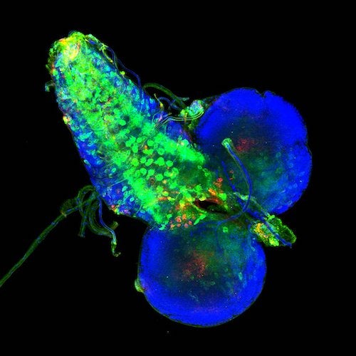
Image courtesy of Abinav Sankaranthi
Determining the spread of tau was a tricky process. Abinav quantified the spread through confocal microscopy, a process that involved mounting the dissected brains of Drosophila larvae onto microscope slides. To pinpoint the location of tau, an antibody treatment was used to stain the tau and green fluorescent protein (GFP) for glutamatergic neurons, which is where the tau originates. The data from confocal microscopy was then uploaded onto the ImageJ software to quantify how much tau was present in the glutamatergic neurons and outside these neurons. They are still not sure how knocking down ITPR and TRPML receptors will affect Tau spreading in the Drosophila brain. According to the images that were taken, the tau proteins seemed to infiltrate the central lobes of the larval brain as well as the regions with glutamatergic neurons.
As a hallmark of AD, the aggregation of tau protein is a complex process with still unknown mechanisms. Now, Abinav is working on fine-tuning the confocal microscopy protocols to obtain more accurate results. He is focused on optimizing his protocol and the quality of his imaging. His work at the Venkatachalam lab will continue to uncover patterns of Tau spread to eventually find a viable target against AD.

